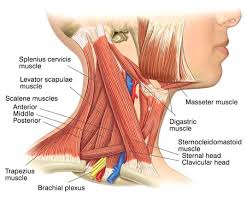
Stiffness in the neck can arise as a result of disorders and diseases of any structure in the neck. The neck contains seven cervical vertebrae that are the bony building blocks of the spine in the neck; these vertebrae surround the spinal cord and canal. Between these vertebrae are discs and nearby pass the nerves of the neck. Within the neck, other structures and organs include the neck muscles, arteries, veins, lymph glands, thyroid gland, parathyroid glands, esophagus, larynx, and trachea. Cervical stiffness is most commonly due to damage to the bones, nerves, and/or muscles of the neck.
Fatigue, like perspiration and aging, is an inevitable feature in the career of any athlete. Fatigue is both a physical and a mental state, representing that point in a difficult training session or competition when the body demonstrates a reduced ability to work efficiently, with a feeling of weariness that cannot be mentally overcome. Fatigue can develop over a short period of time in the course of an event or in practice. It may also slowly develop in the athlete as a cumulative effect after weeks or months of intense physical effort.As fatigue can occur in both training and in competitive situations, it is a condition largely determined by the approach taken by an athlete to workouts. Both the duration of training sessions and their intensity are factors underlying the presence of fatigue. The relationship between workouts and a competitive schedule is also an important consideration. The existence of fatigue, and the ability of the body to overcome its effects, is also closely linked to diet and nutrition, the quality of sleep enjoyed by the athlete, scheduled recovery periods, and external factors such as employment pressures, educational studies, and injury. Fatigue is generally episodic. By addressing the identified underlying causes of fatigue, an athlete can reduce or eliminate its impact upon athletic performance very quickly. When the symptoms of fatigue continue in the face of efforts to eliminate them, the athlete must consider the possible existence of chronic fatigue syndrome, a physical condition that requires a comprehensive medical review. Exercise quality can be assessed using two different yardsticks. The power with which an athlete can perform is closely related to the concept of the intensity of the exercise. The intensity will be tied to such physical factors as the maximum oxygen uptake of the athlete (known as the VO2 max), the power of the heart to propel blood through the body, and muscular strength. The second measure is the work capacity of the athlete—the amount of exercise that can be performed. Using the power / intensity and work / capacity definitions, fatigue presents in some important aspects of athletic performance. Muscle fatigue is the most common kind of reduction of physical capacity. Muscle fatigue is a decrease in the available power coming from the muscle over time, and it arises in three different scenarios. In short distance activities such as sprinting, when the muscle is directed to make a maximum number of muscle contractions in a short period of time, fatigue sets into the muscle when the fast-twitch fibers cannot maintain muscle tension. Through training techniques such as interval training, the muscle fibers are conditioned to respond for longer periods to keep fatigue away from the function for longer periods. In longer activities such as distance running, the muscles are directed to contract more slowly, through the present slow-twitch fibers. In these types of activities, the fatigue will arise through a combined effect of muscle repetition and depleted energy stores. Localized muscle fatigue occurs in sports such as kayaking or distance cycling, when a large muscle group is working on an anaerobic basis and the balance of the body is being powered aerobically; the anaerobically powered muscles will experience fatigue not experienced in the rest of the body. Fatigue has a pronounced impact upon the central nervous system and its functions. A fatigued system will tend to have a heightened sensitivity to cold, as well as a reduced ability to maintain a body temperature to prevent the onset of hypothermia (a potentially fatal cold weather condition in which many systems of the body, including optimal brain function, begin to fail when the body temperature is reduced to 95°F [35°C] or below). Tired sailors who fall into cold water will tend to have their body temperature fall at twice the rate as those who enter the same waters but are otherwise relatively rested. In such circumstances, the thought processes of the tired sailor quickly become impaired, and the ability of this person to make decisions regarding rescue options are much reduced. The effects of fatigue are also demonstrated upon the cognitive and the learning capabilities of athletes in both training and in competition. Coaches seeking to teach a technique to an athlete will have greater instructional success earlier in an intense workout, as the athlete will be able to focus on the teaching message without the interference of the fatigue signals sent by the body. In team sports, instruction to the team is often best reserved for a distinct strategy session as opposed to the practice field for this reason.
Diabetic retinopathy is retinopathy caused by complications of diabetes mellitus, which can eventually lead to blindness. it is an ocular manifestation of systemic disease which affects up to 80% of all patients who have had diabetes for 10 years or more. ... Condition of increased pressure within the eyeball, causing gradual loss of sight A viral disease of horses, symptoms of which include fever, spontaneous abortion, and redness of the eyes Condition of lacking visual perception due to physiological or neurological factors. Condition in which the lens of the eye becomes progressive
Dizziness is one of the most common symptoms that will prompt a person to seek medical care. The term dizziness is difficult to understand since it means different things to different people. It is either the sensation of feeling lightheaded as if the individual is weak and will pass out, or it describes vertigo or the sensation of spinning, as if the affected person just got off a merry-go-round. Lightheadedness is often caused by a decrease in blood supply to the brain, while vertigo may be caused by disturbances of the inner ear and the balance centers of the brain. It is important that the health care professional understand the complaint the person is experiencing. That is the first step so that the proper direction can be taken for a diagnosis and treatment.
If you suffer from migraine headaches, you're not alone. About 12 percent of the U.S. population gets them. Migraines are recurring attacks of moderate to severe pain. The pain is throbbing or pulsing, and is often on one side of the head. During migraines, people are very sensitive to light and sound. They may also become nauseated and vomit. Migraine is three times more common in women than in men. Some people can tell when they are about to have a migraine because they see flashing lights or zigzag lines or they temporarily lose their vision. Many things can trigger a migraine. These include Anxiety
Stress
Lack of food or sleep
Exposure to light
Hormonal changes (in women)
Doctors used to believe migraines were linked to the opening and narrowing of blood vessels in the head. Now they believe the cause is related to genes that control the activity of some brain cells. Medicines can help prevent migraine attacks or help relieve symptoms of attacks when they happen. For many people, treatments to relieve stress can also help.
Headache is pain in any region of the head. Headaches may occur on one or both sides of the head, be isolated to a certain location, radiate across the head from one point, or have a vise-like quality. A headache may be a sharp pain, throbbing sensation or dull ache. Headaches may appear gradually or suddenly, and they may last less than an hour or for several days
Greenstick fracture: an incomplete fracture in which the bone is bent. This type occurs most often in children.
Transverse fracture: a fracture at a right angle to the bone's axis.
Oblique fracture: a fracture in which the break has a curved or sloped pattern.
Comminuted fracture: a fracture in which the bone fragments into several pieces.
An impacted fracture is one whose ends are driven into each other. This is commonly seen in arm fractures in children and is sometimes known as a buckle fracture. Other types of fracture are pathologic fracture, caused by a disease that weakens the bones, and stress fracture, a hairline crack.
Other types of fracture are pathologic fracture, caused by a disease that weakens the bones, and stress fracture.
The severity of a fracture depends upon its location and the damage done to the bone and tissue near it. Serious fractures can have dangerous complications if not treated promptly; possible complications include damage to blood vessels or nerves and infection of the bone or surrounding tissue. Recuperation time varies depending on the age and health of the patient and the type of fracture. A minor fracture in a child may heal within a few weeks; a serious fracture in an older person may take months to heal.
Sprains and strains are used interchangeably to describe everything from a twisted ankle to a pulled hamstring, but they are two specific injuries.
A sprain is a stretch or tear in a ligament (the bands of fibrous tissue that connect our bones at the joints).
A strain is also a stretch or tear, this time affecting the muscle itself or a tendon (the tissue that connect the muscles to the bones).
How Does a Sprain or Strain Occur?Sprains usually happen when a person falls, twists, or is hit in a way that forces the body out of its normal position. The most common type of sprain is a sprained ankle; about 25,000 people sprain an ankle every day. Think of a runner who goes over a curb and catches her foot, twisting the ankle; or a baseball player who slides into home plate and twists his knee. Wrist and thumb sprains are also common, particularly in sports like skiing, where it’s not unusual to fall and land on an outstretched palm. Strains happen when a person twists or pulls a muscle or a tendon. Athletes in contact sports, like football, hockey, and boxing, are at particularly high risk for strains. Frequent repetitive motions, like those used in tennis, rowing, and golf, can lead to strains of the hand and forearm. In the workplace, people at particular risk for sprains and strains are those who engage in frequent heavy lifting, which can include everyone from construction workers and laborers to warehouse workers, nurses, and physical therapists. How Can You Detect a Sprain or Strain?The signs of most sprains or strains are very similar: pain and inflammation, and sometimes bruising, at the injured area. Depending on the severity of the sprain or strain, the pain may be mild, moderate, or severe. The more severe the sprain or strain, the more difficult it is to use the affected area. Someone with a mild ankle sprain may just favor that ankle slightly, while a more severe ankle sprain may cause much more pain and make it difficult or impossible to walk. Sprains are graded on a scale of I to III:
Grade I is stretching of the ligament or a very mild tear, with little to no instability at the joint.
Grade II is a more serious but still incomplete tear.
Grade III is a completely torn or ruptured ligament. This is not a broken bone, but can feel like one since it is often impossible to put weight on the joint or use the affected limb.
Ligament injuries in the knee - such as an anterior cruciate ligament (ACL) -- are dreaded by professional and amateur athletes alike. They can be painful and debilitating. They can even permanently change your lifestyle.
But there's good news. While an ACL or other ligament damage once ended the career of many an athlete, treatment is much more successful now.
So what's behind these feared injuries? Ligaments are tough bands of tissue that connect the bones in your body. Two important ligaments in the knee, the ACL and Posterior Cruciate Ligament (PCL), connect the femur or thigh bone with the tibia, one of the bones of the lower leg. But too much stress on these ligaments can cause them to stretch too far -- or even snap. ACL injury and other ligament injuries can be caused by:
Twisting your knee with the foot planted.
Getting hit on the knee.
Extending the knee too far.
Jumping and landing on a flexed knee.
Stopping suddenly when running.
Suddenly shifting weight from one leg to the other.
Recovery time depends on how severe your knee ligament injury is. Whatever you do, don't rush things. These injuries are common in soccer players, football players, basketball players, skiers, gymnasts, and other athletes. There are four ligaments in the knee that are prone to injury:
Mentioned above, the anterior cruciate ligament (ACL) is one of the two major ligaments in the knee. It connects the thigh bone to the shin bone in the knee. ACL injuries are a common cause of disability in the knee. In the U.S., 95,000 people get them every year. They are more common in women than men.
The posterior cruciate ligament (PCL) is the second major ligament in the knee connecting the thigh bone to the shin bone in the knee.
The lateral collateral ligament (LCL) connects the thigh bone to the fibula, the smaller bone of the lower leg on the lateral or outer side of the knee.
The medial collateral ligament (MCL) also connects the thigh bone to the shin bone on the medial or in side of the knee.
The knee joint is a relatively complex anatomical structure. In addition to a variety of ligaments to maintain stability and the presence of large muscle groups, internally, it is a classic example of a synovial joint. Both the femur and tibia are enclosed in a joint capsule lined with synovial tissue. Between the condyles of the femur and the condylar surface of the tibia are menisci, which serve as shock absorbers for the knee joint, located medially and laterally inside the joint. Between each muscle group are fluid-filled sacs called bursa and the presence of fat bodies named for their location, which reduce friction and lend added protection to the joint capsule.During normal activity such as walking or running, and even for support while standing, the knee will function superbly. It can tolerate moderate stress without significant injury. However, the knee lacks support to withstand many types of injury, especially rotational forces such as those seen in many athletic activities. Knee injuries, even though minor, may require surgery, and if they involve the cartilage, may have delayed healing time due to a lack of blood supply to the cartilage.
The femur, or thighbone, is the longest and largest bone in the human body. At its top, it creates the ball-and-socket joint of the hip; it lower end helps creates the knee joint. The second largest bone in body is the tibia, also called the shinbone. This long bone connects with the knee at one end and the ankle at the other. Next to the tibia is the fibula, the thinner, weaker bone of the lower leg. It is also known as the calf bone, as it sits slightly behind the tibia on the outside of the leg. The fibula is connected via ligaments to the two ends of the tibia. The patella, commonly known as the kneecap, is at the center of the knee. It aids in knee extension and protects the joint. As the knee bends, the patella slides along a groove in the femur. Below the tibia and fibula are seven bones known as the tarsals. These make up the ankle and upper portion of the foot. The seven tarsal bones are:
Calcaneus: The largest bone of the foot, it is commonly referred to as the heel of the foot.
Talus: This bone creates the lower portion of the ankle joint.
Cuboid: This multisurface bone sits on the outside of the foot near the fifth phalange (little toe).
Cuneiforms: These three small bones are closest to the five metatarsal bones. They sit in a row beginning at the inside of the foot and end at the cuboid.
Navicular: This curved bone sits between the talus and cuneiforms.
The five metatarsal bones in each foot create the body of the foot. Numbered one through five, the bone that sits behind the big toe is No. 1 and the one behind the little toe is No. 5. The phalanges make up the toes. Each toe consists of three separate bones and two joints, except for the big toe, which only has two bones and one joint like the thumb in the hand. The three toe bones include the distal phalanges at the tip, middle phalanges, and proximal phalanges closest to the metatarsals. The big toes don’t have middle phalanges
The primary protein that makes up bone, collagen, has a higher tensile strength than steel, but it has a flexibility that allows it to absorb tremendous pressure. A mineral, calcium phosphate, helps create hard bone. Because of this, bones are both strong and flexible. This is important for the large bones in the arm as it is human instinct to throw our arms in front of us during moments of panic, whether it be a car accident or simply bumping into a wall. The large bones of the arm include:
Humerus: This bone runs down from the shoulder socket and joins the radius and ulna at the elbow.
Radius: A forearm bone, it runs from the elbow to the thumb side of the wrist.
Ulna: This forearm bone runs from the elbow to the “pinkie” side of the wrist.
These three bones join to form the elbow. The elbow actually forms three different positions when the heads of the three bones vary slightly. These movements are so small that the untrained eye rarely notices the changes in position. The ends of the radius and ulna join bones at the hand to form the wrist, which is formally known as the carpus. Together with the bones in the palm of the hand, these bones form three rows. The bones of the wrist are:
Including those of the wrist and palm, the hand has 27 bones. Each finger has three series of bones:
Proximal phalanges: The largest of the three, these extend directly from the palm. These are where rings rest.
Intermediate phalanges: These are situated between the two joints of the fingers.
Distal phalanges: These are your fingertips.
Bone fractures are among the most common short-term injuries of the arm. These typically occur during high-impact collisions such as automobile accidents, falls, and sports injuries. A fracture to one of the bones in the wrist is one of the most common bone breaks.
Pivot joints (known also as rotary joints). These joints allow for rotation around an axis. There is a pivot joint near the top of your spine that allows your head to move from side to side.
Hinge joints. This type of joint can open and close like a door. Your elbow is a hinge joint. Your biceps and triceps muscle are basically two people standing on opposite sides of a wall (the humerus, or upper-arm bone), each with one hand reaching over to its respective side of a door (the bones of the lower arm). The biceps "shuts" the door, by contracting and lessening the degree of the joint angle, and the triceps, when it pulls on its respective side of the door, "opens" the door, as the hinge then widens.
Gliding joints (known also as plane joints). This type of joint features two bone plates that glide against one another. The joints in your ankles and wrists are gliding joints. (Holding your forearm steady while your hand points upward and then waving side-to-side with your hand is an example of this joint's functioning.)
Ball-and-socket joints. This is the most maneuverable type of joint. Your shoulder and your hip are both ball-and-socket joints. These joints feature a connection between one bone-end equipped with a protrusion that fits into the receptive space at the end of the other bone in the joint. These joints allow for forward motion, backward motion and circular rotation.
Saddle joints. These joints allow for two different types of movement. For instance, a saddle joint allows your thumb to move toward and away from your forefinger (as when you spread all five digits out, then bring them all together side-by-side) as well as cross over the palm of your hand toward your little finger.
Conyloid joints. These joints are similar to ball-and-socket joints, just without the socket (the "ball" simply rests against another bone end).
The human body is the entire structure of a human being and comprises a head neck trunk (which includes the thorax and abdomen), two arms and hands and two legs and feet. Every part of the body is composed of various types of cell At maturity, the estimated number of cells in the body is given as 37.2 trillion. This number is stated to be of partial data and to be used as a starting point for further calculations. The number given is arrived at by totalling the cell numbers of all the organs of the body and cell types.The composition of the human body shows it to be composed of a number of certain elements in different proportions
The study of the human body involves anatomy and physiology. The human body can show anatomical non-pathological anomalies which need to be able to be recognised. Physiology focuses on the systems and their organs of the human body and their functions. Many systems and mechanisms interact in order to maintain homeostosis.
A Sprainned ankle is where the tendons and ligaments are over stretched. the causes for this are due to not warming up properly. symptoms include severe swelling, when you put the foot on the floor it hurts. usual recovery time for this injury is usually 3 to 4 weeks. if you have a sprained ankle or you need further advice or treatment then contact me and I would be delighted to assist and treat your injury